About Cynthia Kiernan

This author has not yet filled in any details.
So far Cynthia Kiernan has created 427 blog entries.

Giving Your New Glasses a Chance

Eye doctors typically pride themselves on being able to improve someone’s vision through glasses or contact lens prescriptions. Whether it’s a first-time glasses wearer, or someone having either a small or large change in their prescription, we like to aim for that goal of 20/20 vision.

Despite our best efforts, however, correcting vision to 20/20 is not always a positive outcome for the patient. Whether someone will be able to tolerate their new prescription is based on something called neuroplasticity, which is what allows our brains to adapt to changes in our vision.

You or someone you know may have had this happen: Your vision was blurry, so you went to the eye doctor. The doctor gave you a new prescription, but after you received your new glasses, things seem “off.”

Common complaints are that the prescription feels too strong (or even too clear!) or that the wearer feels dizzy or faint. This is especially true with older patients who have had large changes in their prescriptions, since neuroplasticity decreases with age. It is also more likely to […]

2025-02-18T05:00:00+00:00February 18th, 2025|Blog|

Have You Heard About MIGS?

For over 40 years the standard surgical treatment for glaucoma was a procedure called a trabeculectomy.

In a trabeculectomy, the ophthalmic surgeon would make a hole in the wall of eye to allow fluid from the inside of the eye to flow out of the eye and then get resorbed by the blood vessels in the conjunctiva (the mucous membrane that covers the white part of the eye).

This surgery often resulted in a large decrease in the Intraocular Pressure (IOP). Reducing the IOP is the goal of glaucoma surgery because multiple studies show that if you can reduce the pressure the progression of glaucoma slows.

The problem with trabeculectomy is that although it frequently lowers the pressure, it also has a fairly high complication and/or failure rate. This led to some reluctance to perform the procedure unless the glaucoma was severe, or the pressure was very high. As a result of those issues there has been a search during the last 40 years for something that had a lower complication rate and could be more easily deployed […]

2025-02-11T05:00:00+00:00February 11th, 2025|Blog|

Chances Are Good You Have These Mites in Your Eyelids

Demodex folliculorum — often just called demodex —  is a mite that occurs naturally on many people's faces and resides in hair follicles, particularly the follicles of eyelashes. Most of the time, these mites cause no problems whatsoever. However, sometimes an infestation can become particularly parasitic, resulting in unhealthy eyelid margins.  This inflammation of the eyelid is called blepharitis.  Blepharitis can be caused by caused by several things, including allergies, bacterial overgrowth, Rosacea and also by demodex.

Often, diagnosis of mite infestation by your eye doctor can be difficult.  The symptoms can mimic other causes of blepharitis, which is one of the most prevalent diseases we see.

The most common sign of a demodex infestation is a cylindrical cuff or "sleeve" at the base of the eyelash. Symptoms include redness, itching, burning, dry eyes and general discomfort in the eyelid.

The probability of demodex infestation increases gradually with age, with nearly 100% of people having demodex in their eyelashes after age 70.  If there are no symptoms present, nothing needs to be done about demodex, as they are […]

2025-01-29T05:00:00+00:00January 29th, 2025|Blog|

Another Perspective on Texting & Driving

There are many opinions on the topic of texting and driving. The goal of this blog post is to explore the effects on vision during texting.

So, from a visual perspective, why does texting make you more likely to crash? The problem lies in distraction from driving. For example, it takes a fast texter approximately 20 seconds to read and reply to a text. At 55 mph on the highway, a driver glances away from the road for approximately one-third of a mile. When the driver is focusing on their screen, this essentially gives the driver tunnel vision, causing the visual system to essentially use peripheral vision for driving. Your central vision is used to detect depth perception, detail, and colors such as red or green. So when texting, your depth perception, or 3-D vision, is altered and if cars are stopped ahead or closing in rapidly, it's not as easily detected. Colors, such as red brake lights or traffic signals, are not as easily noticed.

Next time you encounter situations with texting and […]

2025-01-21T05:00:00+00:00January 21st, 2025|Blog|

When It Looks Like Your Eye Is Bleeding

We commonly see patients who come in saying that their eyes are bleeding.

The patient is usually referring to the white part of their eye, which has turned bright red. The conjunctiva is the outermost layer of the eye and contains very fine blood vessels.  If one of these blood vessels breaks, then the blood spreads out underneath the conjunctiva. This is called a subconjunctival hemorrhage.

A subconjunctival hemorrhage doesn't cause any eye pain or affect your vision in any way. Most of the time, a subconjunctival hemorrhage is asymptomatic.  It is only noticed when looking at the mirror or when someone else notices the redness of the eye.  There should not be any discharge or crusting of your lashes.  If any of these symptoms are present, then you may have another eye condition that may need treatment.  

What causes a subconjunctival hemorrhage?  The most common cause is a spontaneous rupture of a blood vessel.  Sometimes vigorous coughing, sneezing, or bearing down can break a blood vessel.  Eye trauma and eye surgery are other causes of subconjunctival […]

2025-01-14T05:00:00+00:00January 14th, 2025|Blog|

Your Eye Floaters and You

Do you have floaters in your vision?

Floaters are caused by thick areas in the gel-like fluid that fills the back cavity of your eye, called the vitreous.

Many people, especially highly near-sighted people, often see some number of floaters for a good portion of their lives. Often, these floaters are in the periphery of the vision and may only be visible in certain lighting conditions. The most frequent conditions are when in bright sunlight looking toward the clear blue sky. I know this from personal experience since I have a floater in my left eye that I most often see when swimming outdoors. Every time I turn my head to the left to breathe I see this floater moving in my peripheral vision.

This is totally harmless other than when I’m swimming in the ocean and swear that sudden object in my peripheral vision is a shark bearing down on me. Some people who have floaters are not as lucky– the floater might be in their central vision and is almost constantly annoying, especially when trying to […]

2025-01-07T05:00:00+00:00January 7th, 2025|Blog|

How Eye Pressure Is Measured

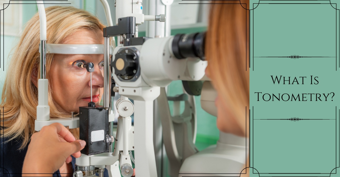
A common question asked during the eye exam is, “When is the puff coming?”  

Patients are referring to air-puff or non-contact tonometry. Tonometry is the procedure used to measure eye pressure, and this is important for diagnosing and monitoring glaucoma.

In non-contact tonometry, a puff of air is used to measure the pressure inside the eye.  The benefit of this test is there is no actual contact with the eye, but the air puff is sometimes very startling for patients. Some people hate that test and it isn’t the most accurate way to measure your eye pressure.

Some doctors don’t even use the air-puff test. Instead, they place a yellow drop that consists of a numbing medicine and then shine a blue light on the eye. This is done in front of the slit lamp and a small tip gently touches the eye to measure the eye pressure. This procedure is called Goldmann tonometry and is considered the gold standard for measuring eye pressure.  

Another method for checking eye pressure is the Tonopen. This is a portable, […]

2025-01-07T05:00:00+00:00January 7th, 2025|Blog|

When the White of Your Eye Turns Red

We commonly see patients who come in saying that their eyes are bleeding.

The patient is usually referring to the white part of their eye, which has turned bright red. The conjunctiva is the outermost layer of the eye and contains very fine blood vessels.  If one of these blood vessels breaks, then the blood spreads out underneath the conjunctiva. This is called a subconjunctival hemorrhage.

A subconjunctival hemorrhage doesn't cause any eye pain or affect your vision in any way. Most of the time, a subconjunctival hemorrhage is asymptomatic.  It is only noticed when looking at the mirror or when someone else notices the redness of the eye.  There should not be any discharge or crusting of your lashes.  If any of these symptoms are present, then you might have another eye condition that could need treatment.  

What causes a subconjunctival hemorrhage?  The most common cause is a spontaneous rupture of a blood vessel.  Sometimes vigorous coughing, sneezing, or bearing down can break a blood vessel.  Eye trauma and eye surgery are other causes of subconjunctival […]

2024-12-31T05:00:00+00:00December 31st, 2024|Blog|

Diabetic Retinopathy ABC’s

Diabetic retinopathy is an eye condition that can affect the retina of people who have diabetes.

The retina is the light-sensitive tissue that lines the back of the eye, and it detects light that is then processed as an image by the brain. Chronically high blood sugar or large fluctuations in blood sugar can damage the blood vessels in the retina. This can result in bleeding in the retina or leakage of fluid.

Diabetic retinopathy can be divided into non-proliferative or proliferative diabetic retinopathy.

Non-proliferative diabetic retinopathy:  In the early stage of the disease, there is weakening of the blood vessels in the retina that causes out-pouching called microaneurysms. These microaneurysms can leak fluid into the retina. There can also be yellow deposits called hard exudates present in the retina from leaky vessels.

Diabetic macula edema is when the fluid leaks into the region of the retina called the macula. The macula is important for sharp, central vision needed for reading and driving. The accumulation of fluid in the macula causes blurry vision.

[…]

2024-12-24T05:00:00+00:00December 24th, 2024|Blog|

Corny Eye Jokes for Your Holiday Get-Togethers

In light of the holiday season, here are our top 10 eye care jokes.

1) What do you call a blind deer? No Eye Deer!

2) What do you call a blind deer with no legs? Still No Eye Deer!

3) Why do eye doctors live long lives? Because they dilate!

4) Why did the blind man fall into the well? He couldn’t see that well.

5) Why shouldn’t you put avocados on your eyes? Because you might get guac-coma!

6) What did the right eye say to the left eye? "Between you and me, something smells."

7) A man goes to his eye doctor and tells the receptionist he’s seeing spots. The receptionist asks if he’s ever seen a doctor. The man replies, “No, just spots.”

8) How many eye doctors does it take to screw in a light bulb? One … or two

9) Unbeknownst to her, a woman was kicked out of peripheral vision club. […]

2024-12-17T05:00:00+00:00December 17th, 2024|Blog|