Blepharitis 101

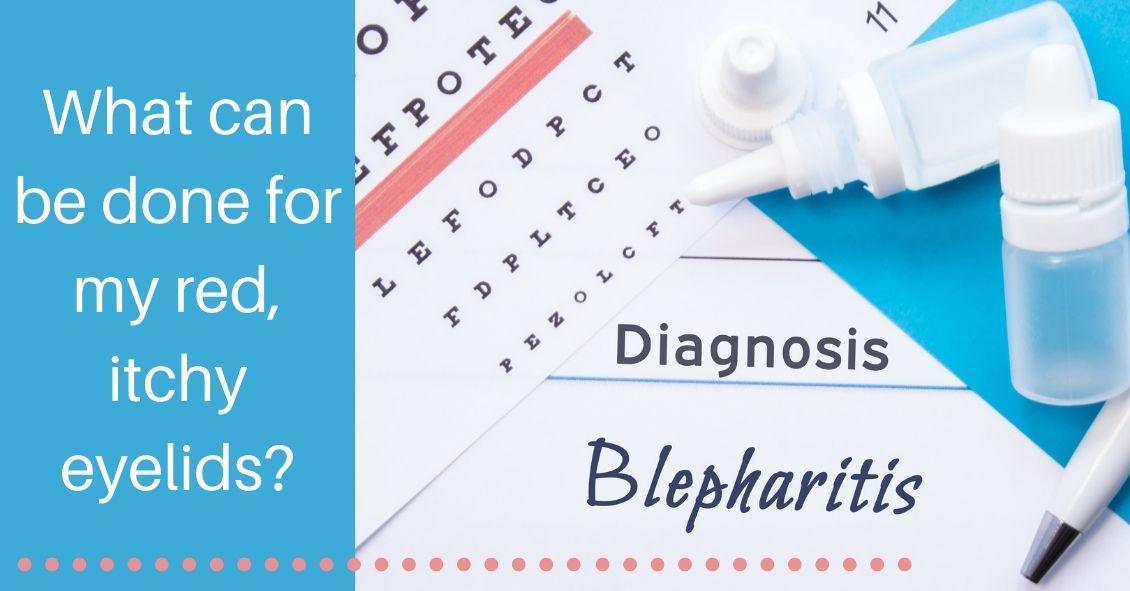
Red, Itchy, swollen eyelids are often due to a condition called blepharitis. Blepharitis tends to be a chronic condition due to thick eyelid mucous gland production that sticks to the bases of the eyelashes. This adherent mucous can allow bacteria to overgrow and also attract and retain allergens. The standard treatment for blepharitis is doing warm compresses and cleaning off the eyelids with a mild baby shampoo and water solution. This treatment works for some people but there are many more sufferers who have chronic irritation and relapses despite this treatment. If the warm compresses and eyelid scrubs are not quite keeping the condition under control there are several other additional treatments that can be used to control the symptoms. One such treatment that your doctor may decide upon is to use an antibiotic/steroid combination drop or ointment. We usually use these for short periods of time to try to bring the condition under control. They are not good to use chronically because it can build resistant bacteria and the steroid component can cause other eye […]

2021-12-14T05:00:00+00:00December 14th, 2021|Blog|

Did You Know You Can Get Freckles in Your Eye?

Choroidal nevus is the fancy term for a freckle in the back of the eye.

This lesion arises from a collection of cells that make pigment in the choroid, which lines the back of the retina and supplies the retina with nutrients. These choroidal nevi (plural of nevus) are usually grayish in color and develop in about 5-10% of the adult population. They are usually asymptomatic and detected during a routine dilated eye exam.

Just like any freckle on our body, we should monitor it for any change in size or growth.  This is usually done with a photograph of the nevus and annual exams are normally recommended to monitor any change.  

In addition to a photograph, other tests that can be used to monitor the nevus are:

  • Optical coherence tomography – a test that uses light waves to take cross-section pictures of the retina. This test is used to detect if the nevus is elevated or if fluid is present underneath the retina.
  • Ultrasound – uses sound waves to measure the size and […]
2021-12-07T05:00:00+00:00December 7th, 2021|Blog|

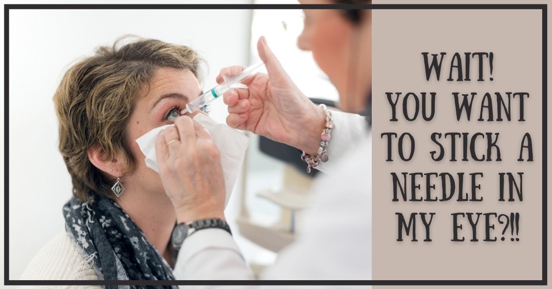
What to Know Before Your First Eye Injection

A common in-office procedure is an eye injection for treatment of various eye conditions, such as wet macular degeneration, diabetic retinopathy, or vein occlusion.  

It is normal to be anxious prior to your first eye injection but knowing what to expect may alleviate some anxiety.  

Your eye will be numbed with topical anesthetic drops. Usually, a cotton-tipped applicator soaked in anesthetic will be held against the white part of the eye to numb it.  Sometimes, a thick gel may also be used for numbing. Once the eye is numbed, it will then be cleaned and a small instrument will be used to hold the eye open. During the actual injection, some patients feel pressure while others may feel a quick sting. Each person will have a different experience and each injection will be different.

The medicine is injected into the white part of the eye. Sometimes a blood vessel may be encountered during the injection and you may notice a little redness on the outside of the eye. This is purely a cosmetic symptom […]

2021-11-30T05:00:00+00:00November 30th, 2021|Blog|

Why Do You Have to Touch My Eye or Puff It with Air?

A common question asked during the eye exam is, “When is the puff coming?”  

Patients are referring to air-puff or non-contact tonometry. Tonometry is the procedure used to measure eye pressure, and this is important for diagnosing and monitoring glaucoma.

In non-contact tonometry, a puff of air is used to measure the pressure inside the eye.  The benefit of this test is there is no actual contact with the eye, but the air puff is sometimes very startling for patients. Most people hate that test and it isn’t the most accurate way to measure your eye pressure.

Some doctors don’t even use the air-puff test. Instead, they place a yellow drop that consists of a numbing medicine and then shine a blue light on the eye. This is done in front of the slit lamp and a small tip gently touches the eye to measure the eye pressure. This procedure is called Goldmann tonometry and is considered the gold standard for measuring eye pressure.  

Another method for checking eye pressure is the Tonopen. This […]

2021-11-23T05:00:00+00:00November 23rd, 2021|Blog|

Winter Is Prime Time for Polarized Sunglasses!

Not everyone understands the importance of sunglasses when the weather turns cold.

Polarized sunglasses are usually associated with Summer, but in some ways it is even more important to wear protective glasses during the Winter.

It’s getting to be that time of year when the sun sits at a much different angle, and its rays impact our eyes and skin at a lower position. This translates to an increase in the exposure of harmful UV rays, especially if we are not wearing the proper sunglasses as protection.

Polarized sunglasses, which are much different than the older dye-tinted lenses, are both anti-reflective and UV resistant. A good-quality polarized sunglass lens will protect you from the entire UV spectrum. This not only preserves your vision, but it also protects the skin around the eyes, which is thought to have a much higher rate of susceptibility to skin cancer.

Snow poses another issue that can be countered by polarized sunglasses.

Snow on the ground tends to act as a mirror because of its white reflective surface and this reflection […]

2021-11-16T05:00:00+00:00November 16th, 2021|Blog|

Diagnosis and Treatment of Diabetic Retinopathy

Diabetic retinopathy, which is a complication of diabetes that affects the eyes, is detected during a comprehensive eye exam that includes:

  • Visual acuity testing.
  • Dilated exam in which drops are placed to widen the pupil to allow examination of the retina.
  • Tonometry. Measurement of the eye pressure inside the eye.

Supplemental testing may include:

  • Optical coherence tomography (OCT). This is a non-invasive test that images the retina to detect any fluid or diabetic macular edema.
  • Fluorescein Angiography. This test involves an injection of a dye into your arm and a series of pictures that are taken as the dye flows through the retinal vessels. This may show leakage of fluid or the growth of new blood vessels in the retina.

Treatment for Diabetic Retinopathy

The best treatment is prevention of diabetic retinopathy by strict control of blood sugars. Once diabetic retinopathy is present, treatment of diabetic retinopathy will slow progression but won’t cure it.

Diabetic macular edema can be treated with several different therapies that may be used alone or in combination.

[…]

2021-11-09T05:00:00+00:00November 9th, 2021|Blog|

Your Vision & Parkinson’s Disease

Parkinson’s disease is a progressive degenerative condition of the neurological system.  The majority of Parkinson’s effects are on movement, often starting off very slowly and subtly. One of the earliest symptoms is a slight tremor in one or both hands. Other early symptoms include a lack of facial expression and decreased blinking of the eyes, so it looks like the person is always staring.  

The next stage usually results in difficulty with initiating movement, especially walking.  It frequently looks like it takes a tremendous concentrated effort to initiate walking and the steps often start off very small with a shuffling of the feet.  At the same time, the disease stiffens the muscles of the arms so that when the person is walking there is a noticeable decrease in the swinging of the arms. Speech becomes much softer and writing becomes more of an effort, with handwriting getting smaller and smaller as the disease progresses.

Parkinson’s can also affect your visual performance, mainly in two parts of your eyes: the tear film and the ocular muscles.

[…]

2021-11-02T04:00:00+00:00November 2nd, 2021|Blog|

Your Eye & Shingles

Shingles is the term we use to describe a condition that is caused by a re-activation of the Herpes Varicella-Zoster virus. The origin of this infection usually goes way back to childhood with a disease we know as chickenpox.

When you have a chickenpox infection your immune system manages to eventually suppress that virus from causing an active infection, but the virus does not get completely eliminated from your body–it is able to go and hide in your nerve roots.

Your immune system manages to keep the virus in check for most of your life but there may come a time in adulthood when your immune system is not working as well as it used to, and the virus can reappear. It usually does this along the distribution of a single sensory nerve called a dermatome.

The most common area for this to occur is along your trunk (chest or abdomen) but it is also commonly found on the face.

There are three branches of nerves that supply sensation to your face. They are all branches […]

2021-10-26T04:00:00+00:00October 26th, 2021|Blog|

What Happened to Those Dilation Reversal Drops?

Getting your eyes dilated can be inconvenient – dilation makes the eyes light sensitive which can make it difficult to read books or use phones and computers for a couple of hours.

So wouldn’t it be great to have another drop to reverse the effects of dilation?

On Dec. 31, 1990, dapiprazole, under the trade name Rev-Eyes, was approved by the FDA and thought to be the answer to all the post-dilation problems. It was marketed for treatment of medically-induced dilation by stimulating pupillary constriction and restoration of accommodative function for reading.

In clinical practice, dapiprazole took between one to two hours to return pupils to pre-dilation size.

Side effects such as stinging upon instillation, conjunctival hyperemia (redness of eye), headache, and a few instances of ptosis (lid drooping), with a possible additional dollar cost to patients, seemed to lessen dapiprazole’s overall clinical benefit.

Reading ability returned in approximately 43 minutes with dapiprazole vs 66 minutes without the drop (Optom Vis Sci 1994 May; 71(5):319-22). The main complaint that people had after using dapiprazole was the […]

2021-10-19T04:00:00+00:00October 19th, 2021|Blog|

What Is Mucus Fishing Syndrome?

The tears that coat the surface of your eyes have both a liquid and a mucous layer to them. It is normal to have a small amount of mucus in your tear film. But that mucus can significantly increase when the eye gets irritated.

Some of the most common causes of irritation that can make the eye overproduce mucus are:

  • Conjunctivitis, which could be caused by an allergy, bacteria, or virus
  • Blepharitis, which is an inflammation of the eyelids
  • Dry Eye Syndrome

When any of these conditions occur, the eye will begin to make more mucus.  

Sometimes the mucous production really is excessive and there is a temptation to keep pulling it out with either your fingers or a cotton swab. DON'T DO THIS–it will just lead to recurring irritation and problems.

Any mucus that gets deposited OUTSIDE the eye on the outer eyelid or on the lashes is fair game for removal. In fact, anything on the exterior of the eyelid or stuck to the eyelashes should be cleaned off.  Just don’t […]

2021-10-12T04:00:00+00:00October 12th, 2021|Blog|